The Bath Vet Referrals surgical team who perform Orthopaedic surgery include Jon Shippam, Cate Khursandi and Edward Corfield.
Orthopaedics
Orthopaedic cases seen at Bath Vet Referrals include lameness investigations and surgical treatments for bone, ligament or tendon injuries and abnormalities.
A lameness investigation consists of taking a detailed history, observing the patient walking to assess lameness and gait, followed by an in-depth physical examination including palpating for evidence of swelling or muscle wastage, looking for signs of pain, assessing range of motion, joint stability and feeling for crepitation. Basic neurological tests are performed to assess for delays in conscious proprioception. In many cases the cause of lameness can be localised to specific areas which allows further investigations such as advanced imaging to help make a diagnosis. Where a diagnosis has already been made we will perform a thorough assessment to confirm the diagnosis and to rule out any concurrent issues.
The method of advanced imaging we most commonly use for orthopaedic conditions is computed tomography (CT). This uses x-rays to create a three-dimensional image that is especially good for identifying bony lesions. Magnetic resonance imaging (MRI) is also useful in some cases - especially if a lameness is actually caused by a neurological problem such as lumbosacral stenosis. Simple fractures, hip dysplasia and cruciate ligament ruptures are examples of conditions where radiography provides sufficient diagnostic information.
We encourage physiotherapy and hydrotherapy in the treatment and post-operative rehabilitation of orthopaedic cases. Our ACPAT physiotherapist is available to provide treatment for post-operative inpatients as well as follow up appointments for outpatients.
Fractures
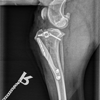
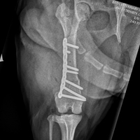
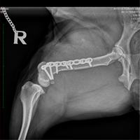
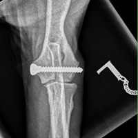
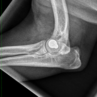
Fractures classifications describe whether they are open or closed, simple or comminuted, displaced or non-displaced and articular or non-articular. We offer a range of fixation methods such as using bone plates (including use of Synthes LCP which allows use of locking internal fixation with the benefits of achieving fracture compression), external skeletal fixation (using the Imex SK Linear ESF System), hybrid fixation (using the Imex SK Hybrid ESF System), Ilizarov ring fixators, and for some avulsion fractures using pins and tension band fixation. Each case is classified and carefully assessed to allow us to recommend the most appropriate method of fixation. Autogenous cancellous bone graft is used to help speed the repair in many cases.
Cruciate ligament ruptures
At Bath Vet Referrals we offer TPLO, TTA, CCWO and lateral suture as treatment options for cranial cruciate ligament (CCL) disease. Cranial cruciate ligament tears or ruptures are most commonly a degenerative process that leads to instability of the stifle (knee) and often causes secondary tearing of the meniscal cartilages. Various methods of treatment are available, but in larger dogs we usually recommend the tibial plateau levelling osteotomy (TPLO) or tibial tuberosity advancement (TTA) procedure (using genuine Synthes and Kyon implants respectively). The TTA and TPLO procedures change the biomechanics of the stifle to overcome the instability caused by failure of the cranial cruciate ligament. We also offer lateral suture stabilisation which can be used in any size of dog but is best suited to small dogs and cats. Cranial closing wedge osteotomy of the tibia (CCWO) is another option similar to TPLO that is recommended in selected cases.
Patellar luxation
Patellar luxation is a common condition that can be asymptomatic through to causing severe mobility issues. The most common presentation is where dogs show intermittent skipping lameness, and often isn’t painful at the time of diagnosis. In the majority of cases the patella (knee cap) luxates because of a malalignment in the hindlimb. Treatment options include compensating for the malalignment by moving the part of the tibia called the tibial tuberosity, or by directly correcting the malalignment by use of a corrective osteotomy which involves straightening the bone (and/or correcting torsion) and using a bone plate to stabilise the bone whilst it heals. Many cases also require deepening of the groove that the patella sits in to improve stability. Studies have shown patellar luxation to have disappointing complication rates including recurrences, but modern techniques including CT alignment measurements can help make a tailored plan for each case which can significantly reduce the complication rates.
Arthroscopy and Elbow Dysplasia
Our facilities include high definition video endoscopy facilities that allow us to perform arthroscopy procedures to both assess and treat conditions inside joints. The most common use of arthroscopy is to assess cases of elbow dysplasia, especially if they have fragmentation of the medial coronoid process. In cases of suspected elbow dysplasia we will usually start with CT, and particularly if this shows fragmentation then we may recommend arthroscopic (key hole) removal of the fragment. In some cases with persistent lameness, loss of cartilage in the medial compartment of the elbow but intact cartilage in the lateral compartment we may recommend the PAUL procedure (proximal abducting ulnar osteotomy) which aims to reduce load on the medial compartment.
Humeral intracondylar fissures (HIF) in Spaniel breeds
Sometimes referred to as incomplete ossification of the humeral condyles, this condition is mainly seen in Spaniel breeds and may be a developmental abnormality but evidence shows some cases develop fissures after being previously documented as normal. CT is the most sensitive method of diagnosing this condition. HIF can cause a progressive forelimb lameness, but most seriously can lead to spontaneous elbow fracture (including lateral condylar fractures or T or Y fractures). The lameness and risk of fracture can be resolved in most cases by placement of a transcondylar screw.
Other conditions and treatments at Bath Veterinary Referrals include:
- Hip luxations
- Shoulder injuries
- Meniscal tears
- Limb deformities
- Immune mediated polyarthritis
- Osteochondrosis dissecans (OCD)
- Stem cell therapy
- Platelet therapy
- Hock and carpal injuries including pancarpal and pantarsal arthrodesis
To refer a case, please click here.
If you would like advice on an Orthopaedic case, please don’t hesitate to contact us.
Meet our Vets

Dr Jonathan Shippam Dr Edward Corfield Dr Cate Khursandi