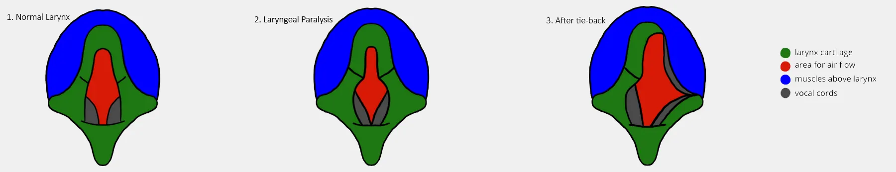
What is the larynx?
The larynx (voice box) is a cartilage structure in the neck of dogs and cats and has three main functions:
- Vocalisation
- Closing the entrance to the airway during swallowing. This prevents food or fluids from entering the airways
- Controls airway resistance – by opening the larynx, more air can flow into the airways during exercise or times of stress.
What is laryngeal paralysis?
Laryngeal paralysis is a loss of movement of the cartilage inside the larynx. During normal breathing, the cartilages move in and out with inspiration and expiration. In laryngeal paralysis, the cartilages can no longer open (and are sometimes sucked inwards) during inspiration. This results in an increase in airway resistance through the larynx and difficulty breathing.
CONGENITAL LARYNGEAL PARALYSIS – this is seen in dogs under 1 year of age and is rare. Most dogs present with an episode of respiratory distress and respiratory noise on exercise or excitement.
AQUIRED LARYNGEAL PARALYSIS – this is the most common form of the disease and is most often seen in middle aged to older large breed dogs (especially Labradors, Golden Retrievers, Setters, St Bernards and Collies). It can be associated with chronic endocrine diseases (especially hypothyroidism) although the concurrent diagnosis of these two conditions is often incidental. Acquired laryngeal paralysis is caused by an abnormality in the nerves supplying the larynx and can often be seen as part of a polyneuropathy complex (other nerves are affected too). Occasionally laryngeal paralysis can be caused by trauma, tumours and previous surgery.
Clinical signs of laryngeal paralysis
The most common clinical sign is a roaring noise when breathing (especially exercising, panting, hot weather and excitement). The onset of this noise is often over a period of months to years with increasing severity and inability to exercise. Owners can often think that the change in breathing and exercise are “normal for old age” due to the gradual onset of clinical signs. Owners will often notice a change in the pitch of the bark, gagging when eating and a harsh cough. During hot weather, dogs with laryngeal paralysis can collapse and even die. This is due to increased panting, inability to lose heat, increased body temperature, airway swelling and anxiety.
Dogs with laryngeal paralysis are more prone to pneumonia secondary to inhalation of food particles and can have additional difficulty breathing due to this.
If laryngeal paralysis is part of a polyneuropathy complex other clinical signs may be present. For example, scuffed claws on the hindlegs, difficulty rising and muscle loss in the limbs. Surgical treatment of laryngeal paralysis will not improve these other clinical signs.
How do we diagnose laryngeal paralysis?
A provisional diagnosis is made from hearing the typical “stridor” noise on clinical examination but a definitive diagnosis cannot be made until the larynx has been directly visualised under a light plane of anaesthesia. The larynx cartilage is checked and if there is lack of movement when the dog is breathing the diagnosis of laryngeal paralysis is made.
Other tests which we may do:
- Blood tests – if these have not recently been performed we would recommend a full blood test including thyroid values. If there is concurrent thyroid disease we would usually treat this before surgery as occasionally laryngeal paralysis signs can improve with medical management of hypothyroidism.
- Checking the gag reflex – this is the reflex at the back of the throat which enables us to check swallowing function.
- Radiography (X-rays) – these will be performed under the same anaesthetic as diagnosis and surgery for laryngeal paralysis. We look for any signs of pneumonia on the radiographs and if this is present we would treat this before surgery.
- CT scan – if we were concerned about any other lung changes a CT scan may be recommended.
Treatment options
Occasionally medical treatment can be successful. This involves weight loss, exercise restriction, anti-inflammatory drugs and avoiding stressful or exciting situations. As the disease is progressive, medical treatment may only be successful for a short period before severe respiratory distress occurs.
Surgical management is more successful and involves placing a permanent suture to hold the larynx open on one side in a procedure called a “tie-back”. This increases the size of the airway enough to improve clinical signs even though only one side of the larynx is held open. The surgery will improve the roaring noise when breathing but often causes a worsening in coughing. If coughing after eating is the main presenting clinical sign (with mild noise) we may recommend medical treatment instead. Surgical management will change the character of the bark.
Post-operative care
Hospitalisation following tie-back procedures is routine for 12-48 hours following the surgery. This enables us to provide post-operative pain relief and to monitor the dog’s breathing for any signs of distress.
Following tie-back procedures a course of antibiotics and anti-inflammatory medication will often be prescribed.
Feeding is particularly important; the dog should be encouraged to eat slowly and fed a diet of sausage meat consistency rolled into meat balls. During hospitalisation we will monitor for signs of coughing when eating. As the larynx is being held open it is easier for the dog to inhale food when swallowing which could cause pneumonia.
Only water should be offered to drink indefinitely. If gravy or milk are inhaled there is a large risk of pneumonia.
Exercise: no exercise is recommended for 2 weeks following surgery. Following this the exercise should be gradually increased. We would recommend using a harness instead of a collar following tie-back surgery. Swimming is not recommended following tie-back surgery.
Barking should be discouraged as this may cause the suture to break. If the dog is barking during hospitalisation we may choose to send the patient home quite soon following surgery.
Complications following surgery
During surgery, fracture of the cartilage can occur when the suture is placed. The tie-back would then be performed on the right side instead.
Acute swelling of the airway in the immediate post-operative period. Although this is rare it is a concern and we will hospitalise dogs following this surgery to monitor for any difficulty breathing.
Fairly commonly a seroma (fluid-filled pocket) forms at the surgical site. This is managed conservatively and will often resolve with time.
Following surgery, the pitch of the bark will change. 10-20% of dogs having a tie-back procedure will have an episode of aspiration pneumonia in the weeks to years following surgery. This often occurs due to the inability to protect the airway during swallowing and inhalation of food particles. The diet is particularly important in preventing this from occurring.
Persistent coughing and gagging is common following surgery. This can improve in the weeks following surgery but often persists long-term.
Recurrence of clinical signs can occur if the sutures we have placed fail. This can be at any time following surgery and can be caused by excessive barking. In these cases, repeat surgery would be required to alleviate the clinical signs.
If you have any questions about this information please raise them during your consultation with the vet.